Abstract
Introduction:
Primary Mediastinal Large B-cell Lymphoma (PMBCL) is one of the primary extranodal lymphomas and originating from the thymic medulla B-cells. PMBCL is localized in the anterior and upper mediastinum, often with compression of the superior vena cava and with tumor infiltration of the pericardium, lungs, pleura. Sometimes there is impairment of the kidneys, adrenals, liver and central nervous system. Usually the bone marrow is not including. According to the WHO Classification 2008, if bone marrow is infiltrated by large B-cell lymphoma with mediastinal tumor mass, differential diagnosis between diffuse large B-cell lymphoma (DLBCL) andPMBCLshould be performed.
Evaluation of the expression level of the genes JAK2, PDL1, PDL2, MAL, TRAF1 by polymerase chain reaction allows to different PMBCL and DLBCL. Diagnosis the PMBCL is established if detection of hyperexpression two or more genes.
We did not find any publications with description of PMBCL with bone marrow involvement. We present here 2 clinical cases.
Materials and methods:
154 patients with PMBCL treated in our center were examined. 16 (10,4%) of them had an extra-mediastinal localization. 2 patients (1,3%) were diagnosed with PMBCL with bone marrow involvement.
Results:
Case 1
A 42 years old female was hospitalized for vital indications with respiratory failure and syndrome of compression of the superior vena cava. In the mediastinum a tumor was 138x116.5 mm, involving the upper and middle lobes of the right lung. Tumor infiltrated the soft tissues of anterior wall of the chest, pericardium.
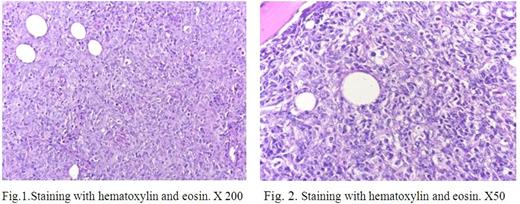
The biopsy of the mediastinal tumor revealed the fragments of fibrous fat tissue with a dense infiltrate of medium size and large cells with oval nuclei, with one large or smalls nucleoli, a moderately pronounced light cytoplasm (Fig.1). Tumor cells expressed the CD20, PAX5, co-expressed CD23, CD30-, IgM-, Ki-67 80%. LDH level in blood was 578 E/l (208-378 E/l). International Prognosis Index (IPI) - 4. Peripheral blood was normal.
Histology of the bone marrow showed diffuse interstitial infiltration by lymphoid cells of medium and large size with several small distinct nucleoli, mainly with the morphology of centroblasts, increased mitotic activity (Fig. 2). Tumor cells of bone marrow were CD20 +.
Hyperexpression of JAK2, PDL1, TRAF1 genes in the bone marrow was revealed by PCR method. So the PMBCL was diagnosed.
The patient received 6 courses of R-DA-EPOCH-21. At the end of treatment tumor lesions were still positive by PET/CT (Deauville score 5). The patient received the second line therapy, with HSCT. PET/CT was positive again (Deauville score 5). Hyperexpression of JAK2, PDL1, TRAF1 genes persisted in the bone marrow. The patient is alive now at 18 month from start of treatment. According to the CT there is no signs tumor progression.
Case 2
B 69 years old female complained of weakness, profuse sweating, pain in the left scapular, temperature 38,0 C, weight loss by 11% of body weight, for the last 3 months.
In the left hemithorax tumor mass of 135x67 mm, embracing the first rib with a pathological fracture. Tumor cells expressed CD20, PAX5, CD79α, TNFAI2, co-expressed CD23 and CD30, Ki-67 - 80%. LDH level in blood 994 E/l. IPI - 4. Peripheral blood was normal.
Histological study of the bone marrow revealed a focal-interstitial lymphoid infiltration with small and medium sized cells with oval and irregularly shaped nuclei. Hyperexpression of the genes JAK2, MAL, TRAF1 was revealed in the bone marrow.
The patient received 6 courses of R-DA-EPOCH-21. After the end of treatment FDG-PET/CT was positive (Deauville score 5). The patient received the second line therapy. After the treatment disease progression was established. Hyperexpression of JAK2, MAL, TRAF1 genes persisted in the bone marrow. The patient is alive now at 18 month from start of treatment, but with PMBCL.
Conclusion:
Both cases demonstrated resistant to R-DA-EPOCH lymphoma, though the frequency of complete remissions in patients with PMBCL is almost 90%. New approadies should be developed in such cases.
Each year, with the accumulation of research data it became evident that within each well-defined nosological form there are borderline cases or those in which our understanding is insufficient to explain a particular phenomenon. The detection of bone marrow involvement in patients with PMBCL makes one think about the criteria for diagnosing the disease.
No relevant conflicts of interest to declare.
Author notes
Asterisk with author names denotes non-ASH members.


This feature is available to Subscribers Only
Sign In or Create an Account Close Modal